Examining and medicating a dog’s eyes

If your veterinarian has prescribed medication for your dog’s eyes, don’t worry – with a little patience it can an easy task.
Medicating the eyes can be messy so cover good clothes and work on a surface that is easy to clean. Some dogs will happily sit in your lap or on a table while you medicate their eyes, but many require some form of restraint.
How to restrain a dog while you examine or medicate its eyes
One method to restrain the dog is to place it on a table. Stand on the side of the table opposite to the eye you are medicating (in the photograph the right eye is being medicated).
Drape your right arm over the dog’s shoulders. Use your left hand to firmly push the dog’s muzzle to the table and to pull the lower eyelid down. Use your right hand to hold the medication container.
If the dog tries to stand, lean your upper body over its shoulders to prevent it from rising.

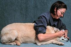
If your dog is too wiggly, try laying it on its side.
Use your right arm and upper body to keep the dog laying on its side. Hold the medication container in your right hand. Use your left hand to keep the head on the table and to pull the lower eyelid down.
It is easier to perform this procedure if you have a helper.
Examining a dog’s eyes

To examine the eyes, the head is cupped between both hands with one thumb on the upper eyelid and the other thumb on the lower eyelid.

To see the parts of the eye beneath the upper eyelid, pull the upper eyelid up with your thumb to open the eye widely. The white part of the eye is the sclera, which is normally glistening white and has small, thin red blood vessels on its surface.
Abnormal findings on the sclera include:
- large, engorged blood vessels
- bruises, which may indicate a local injury or a problem with the clotting system
- yellow discoloration of the sclera, which indicates jaundice
If you stretch the lid more, you will see a pink tissue, which is the conjunctiva. In health, the conjunctiva should be about the same shade of pink as the gums.
Abnormal findings on the conjunctiva include:
- pale pink, which may indicate anemia
- yellow discoloration, indicating jaundice
- bruises, which may indicate a local injury or a problem with the clotting system
Peering through the pupil, you look through the lens, which is clear, and you may see a bright colorful structure, which is the retina.
The iris can be one of several different colors and some dogs have two different colors. Some, but not all, dogs with blue eyes are deaf.
Abnormal findings on the iris include:
- ragged edges, although this can occur with aging and is called iris atrophy
- growths on the iris
- black spots on the iris
- blood spots on the iris
The pupil is the black spot in the center of the eye. Dog pupils are round. The pupils should be the same size and should constrict to a pinpoint when a bright light is shined in the eye. The pupil is a hole in the center of the iris. The lens is behind the pupil but is not seen when healthy, as it is clear.
Abnormal findings in the pupil include:
- cloudy or blue discoloration of the pupil, indicating cataracts or an aging change called nuclear sclerosis
- different sized pupils, which is called anisocoria
- ragged edges, although this can occur with aging


Use your lower thumb to pull down the lower eyelid. When you pull the lower lid down it pulls away from the eyeball and creates a pouch that is lined by pink conjunctiva. This pouch is where eye medications are placed.
When you pull down the lower lid you may also see the third eyelid, also called the nictitating membrane, that will protrude over the bottom inner corner of the eye. The third eyelid is usually a pale pink or white color and has thin blood vessels on its surface. The third eyelid is not visible in the above photograph.
Abnormalities of the conjunctiva and third eyelid include:
- pale pink, which may indicate anemia
- yellow discoloration, indicating jaundice
- discharge accumulation in the pocket
How to administer eye medication to a dog

Eye medications are either drops or ointments. Ointments stay in the eye longer than drops and are usually applied less often. Your veterinarian will prescribe specific medications for specific conditions.

Cradle the head in one hand, usually the left hand if you are right-handed. Use the thumb of the hand holding the head to pull down the lower eyelid to create a pouch. Hold the ointment tube in your right hand. With the tip a few millimeters away from the eye, not touching the eye, squeeze a small ribbon of ointment into the pouch.

To distribute the ointment across the eye, massage the ointment across the surface of the eye with eyelids closed.

Eye drops are also placed in the pouch created when you pull down the lower eyelid. Hold the head and pull down the lower eyelid as described for placing ointments. Drop the prescribed number of drops into the pouch without the tip of the bottle touching the eye. Eye drops disperse across the surface of the eye rapidly and do not need to be rubbed across the eye by massaging.
Depending upon the size of the dog’s head and your hands, you may rest the middle finger or heal of the hand holding the bottle or tube on the dog’s head to keep your hand steady and reduce the risk of poking the dog in the eye with the bottle or tube.
This information is not meant to be a substitute for veterinary care. Always follow the instructions provided by your veterinarian. Washington State University assumes no liability for injury to you or your pet incurred by following these descriptions or procedures.